sdfsdfsdf
What are Complex Lymphatic Anomalies?
CLAs are a group of rare developmental diseases characterized by abnormal growth of lymphatic vessels. They may involve multiple organ systems, including the lung, spleen, soft tissue, and bones. CLAs can manifest anywhere in the body and are associated with severe morbidity and health complications, including pain, swelling, infection, and lymphatic leakage (Reference 1).
CLAs include:
- Gorham Stout Disease (GSD)
- Generalized Lymphatic Anomaly (GLA)
- Kaposiform Lymphangiomatosis (KLA) and
- Central Conducting Lymphatic Anomaly (CCLA).
CLAs have a broad spectrum of symptoms and phenotypes. Consequently, patients with the same diagnosis may have different symptoms based on the location of the body that is involved.
It is essential to promote awareness of CLAs, provide resources and support to patients, and assist clinicians in gaining more information about these rare diseases to help patients reach an earlier diagnosis and receive optimum care.
For more information on the classification of CLAs, you can visit the International Society for the Study of Vascular Anomalies (ISSVA) (Reference 2).
The Lymphatic System
To understand CLAs and their manifestations, one must understand normal lymphatic structure and function.
The lymphatic vasculature is a unidirectional conducting system that returns filtered interstitial fluid and tissue metabolites to the blood circulation and plays major roles in immune cell trafficking and lipid absorption. Initial lymphatics consist of a single layer of loosely connected lymphatic endothelial cells (LECs) and are responsible for the uptake of excess fluid and macromolecules in tissues.
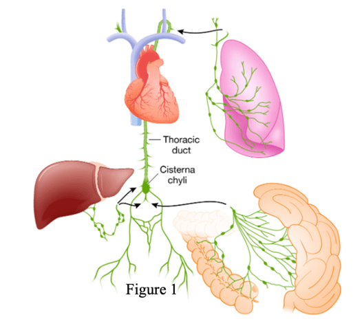
Initial lymphatics first drain into pre-collecting lymphatic vessels that merge with larger collecting lymphatics. The smaller lymphatic vessels, which take up the fluids, are called lymph capillaries. The lymphatic capillaries are composed of a single layer of endothelial cells (LECs). They drain into collecting lymphatics, which are distinguishable by the presence of a smooth muscle layer and one-way bicuspid valves to prevent retrograde fluid flow. Eventually the collecting lymphatics throughout the body coalesce into the larger lymph trunks, of which the largest, the thoracic duct and right lymph duct, empty directly into the subclavian veins.
The lymph from the lower limb terminates at the para-aortic nodes. They join with the lymph from the viscera of the pelvis and form bilateral lumbar trunks. The lumbar trunks, hepatic and intestinal lymphatics drain into the cisternae chyli, a structure that marks the beginning of the thoracic duct. Anywhere from 50% to 90% of lymphatic fluid is derived from the intestine and liver. Intestinal lacteals contain mostly dietary fat, and ingesting a fatty meal can increase the lymph flow in the lacteals ≥200-fold compared with a fasting state.
Important parts of the lymphatic system are:
- Lymph: Clear fluid containing white blood cells that helps clear toxins and waste
- Chule: A milky fluid consisting of fat droplets and lymph. It drains from the lacteals of the small intestine into the lymphatic system during digestion
- Lymphatic vessels: Small tubes (vessels) that carry lymph throughout the body.
Genetics of CLAs
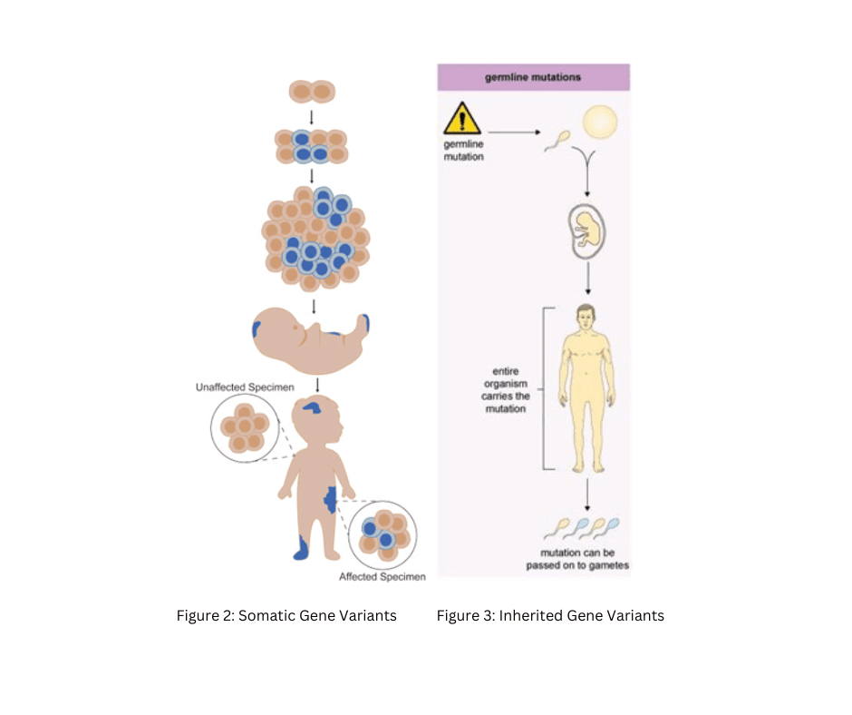
Vascular anomalies are caused by post-zygotic (somatic) (Figure 2) or germline (Figure 3) pathogenic variants in genes that regulate cell growth and vascular development (Figure 4). The clinical variability seen in CLAs is believed, in part, a reflection of the somatic nature of these driver mutations and is highly dependent on when the variant occurs during development, where the affected cells and tissues are located, which specific alleles are present, and how the variant affect the function and expression of the altered genes (Reference 3).
Acquired somatic variant or mutation: An alteration in DNA that occurs after conception. Acquired somatic variants can occur in any of the cells of the body except the germ cells and therefore are not passed on to children. (Figure 2)
Germline (or inherited) variant: A DNA alteration within a germ cell during conception. Germline variants therefore become incorporated into the DNA of every cell in the body. Germline variants are passed on from parents to offspring. (Figure 3)
The majority of CLAs are caused by acquired somatic gene variants